Watching materials grow
My research focused on how materials form and how their structure affects their properties.
I earned my PhD in Materials Science and Engineering at National Chiao Tung University, now part of National Yang Ming Chiao Tung University, in 2016. My advisor was Prof. Ying-Hao Chu, and my co-advisor was Dr. Haimei Zheng.
From 2012 to 2016, I was a visiting scholar in Haimei Zheng’s group at Lawrence Berkeley National Laboratory. There I used liquid-cell electron microscopy, which seals a thin layer of liquid inside the microscope so crystals can be filmed as they grow.
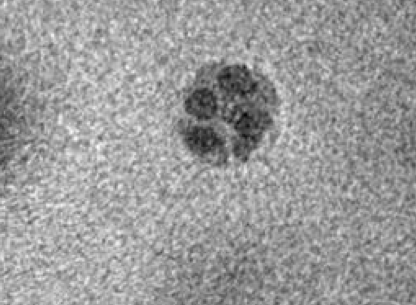 t = 0.1 s
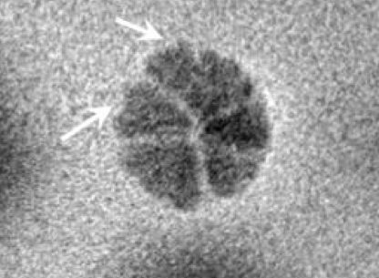 t = 29.8 s
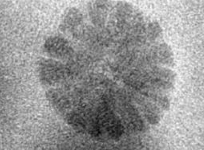 t = 75.7 s
Some crystals grow into branches, called dendrites. They also form on battery electrodes, where they damage the cell.
I filmed iron oxide and nickel ferrite dendrites from the first nanoparticles to fully branched structures. A tip widens before it splits, and faster-growing tips split wider.
Related paper: Nano Letters, 2018
The first question was which metals end up in the crystal. Nickel and cobalt grew into the crystals with iron, while manganese and zinc stayed in the solution. The outcome followed how easily each metal is reduced and how readily its starting compound breaks down.
I then mixed platinum with iron. With four times more iron than platinum, an iron–platinum core formed first. When the platinum ran out, an iron oxide shell grew around it. We recorded the process at atomic resolution.
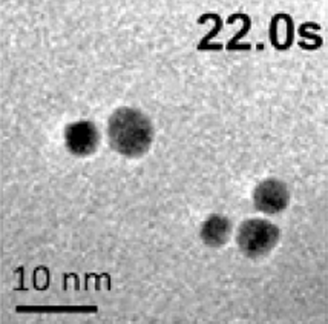 t = 22.0 s
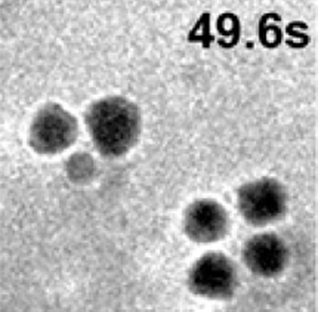 t = 49.6 s
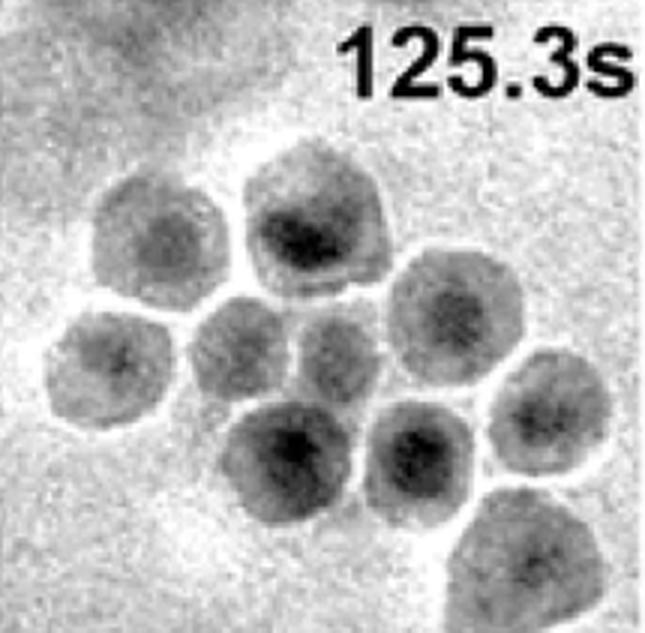 t = 125.3 s
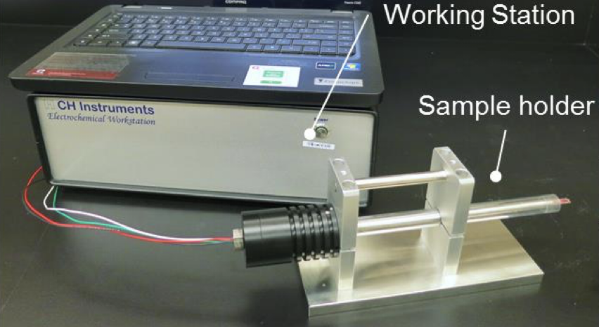
To run electrochemistry experiments inside the microscope, I built a new sample holder. The group’s earlier holder had two electrodes and needed wire bonding for each experiment, which often broke the liquid cell.
With Haimei Zheng’s guidance, I designed a three-electrode holder for FEI microscopes, drew the 3D model, and worked with a machine shop to CNC-machine the parts. In testing, it gave a much steadier current signal than the earlier holder.
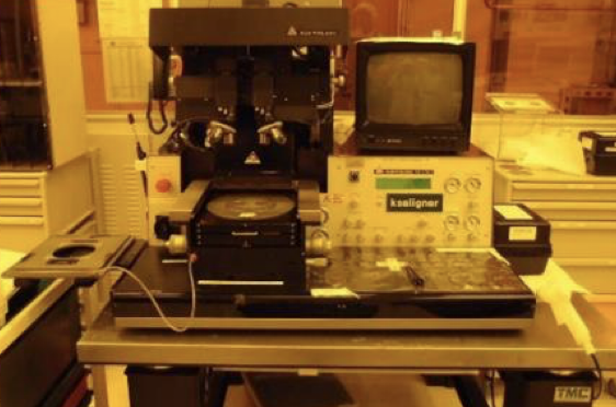
I made the liquid cells at the Marvell Nanofabrication Laboratory at UC Berkeley, working through lithography, etching, and thin-film deposition on silicon wafers. Some cells carried electrodes, so we could watch electrochemical reactions, including battery charge and discharge.
As the only PhD student in the group at the time, I contributed hands-on work across several projects.
Before Berkeley, I spent about three years growing oxide thin films in Hsinchu, mainly BiFeO₃ and self-assembled oxide nanocomposites, using pulsed laser deposition. I also grew samples by molecular beam epitaxy for collaborators.
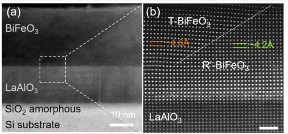
BiFeO₃ can form a nanoscale boundary between two crystal phases, where its electrical and magnetic response is enhanced. That boundary had been made on oxide substrates. We made it on silicon, a step toward using it in semiconductor devices, and I mapped how the phases develop as the film gets thicker.
The research resulted in 29 journal papers, including four first-author papers.
I also co-authored three papers in Nature Communications and three in Nano Letters.
Google Scholar, October 2026: 2,044 citations · h-index 22 · i10-index 25
Selected first-author papers
- In situ study of Fe₃Pt–Fe₂O₃ core–shell nanoparticle formationJournal of the American Chemical Society, 2015
- In situ study of spinel ferrite nanocrystal growth using liquid cell TEMChemistry of Materials, 2015
- Epitaxial integration of a nanoscale BiFeO₃ phase boundary with siliconNanoscale, 2016
- Design of magnetoelectric coupling in a self-assembled epitaxial nanocomposite via chemical interactionJournal of Materials Chemistry C, 2014
PhD thesis
- The Growth Kinetics Study of Functional Nanostructures using In-situ Transmission Electron Microscopy in SolutionNational Chiao Tung University